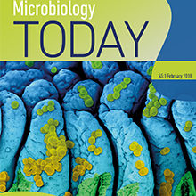
Microbiology Today February issue: Imaging
13 February 2018

The first issue of Microbiology Today for 2018 focuses on the variety of ways that humans have been able to view micro-organisms. Beginning with the microscope – most famously used by Antonie van Leeuwenhoek to study ‘animalcules’ – our feature articles demonstrate some of the cutting edge methods of showing microbes in closer, and higher quality, detail than ever before.
Pippa Hawes introduces us to The Pirbright Institute’s unique bioimaging facility, which is helping scientists understand how deadly livestock viruses function to improve diagnoses, vaccine research, and outbreak predictions. In the next article, Christopher Bartlett points out how a higher level of magnification is not enough to achieve good, crisp image resolution, due to light diffraction. Christopher discusses how using a type of super-resolution microscopy called ‘single molecule localisation microscopy’ (SMLM) can, not only bypass the issue, but can also be used as a quantitative tool.
Next, Jessica Mark Welch considers the difficulties of studying microbes in their natural habitats, in particular the diverse communities of bacteria that might look near-identical under a microscope. The solution, she explains, is fluorescence in situ hybridisation (FISH), which allows researchers to see different types of bacteria by making them glow. Gail McConnell, Brad Amos and Liam Rooney explore the possibilities of the Mesolens imaging system, a giant lens that can resolve individual bacteria in a much larger volume than was previously possible. Originally designed for imaging rodent embryos, Gail, Brad and Liam believe the Mesolens has the potential to have much broader applications.
Michele C. Darrow and Karen E. Marshall discuss using X-rays to study disorders associated with protein misfolding, like Alzheimer’s disease and Huntington’s disease. Although using X-rays has considerably decreased the amount of time needed to visualise what is occurring at the cellular machinery level, Michele and Karen explain that more could still be done to improve how long it takes. The Comment piece from Bruno Martins and James Locke outlines how time-lapse imaging can bring single-celled organisms to life, and how these movies can help researchers understand both how and why microbes do the things they do.
Within this issue are also updates from our journals, details about the upcoming Annual Conference in April, outreach activities run by Society members, and more.
View the latest issue online.
