In pictures: The first images of a coronavirus
Posted on April 17, 2020 by Laura Cox
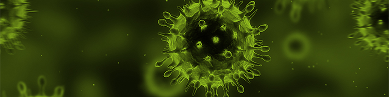
Today, the word ‘coronavirus’ has become a part of everyone’s daily vocabulary. It’s difficult to believe that 60 years ago the very existence of these viruses was disputed by experts, with the first images of coronavirus written off as badly imaged influenza. Now, as the coronavirus SARS-CoV-2 continues to change the world as we know it, the history of coronaviruses is being revisited.
First described by virologists June Almeida and David Arthur John Tyrrell in 1964, the first microscopy images of human coronaviruses were published in the Journal of General Virology two years later.
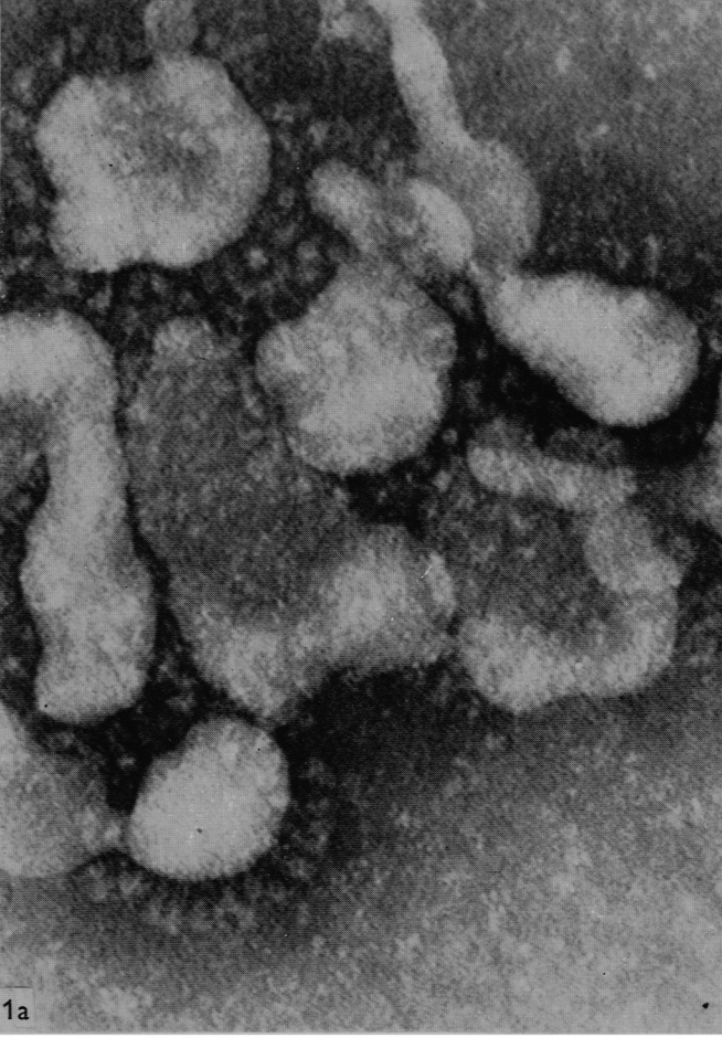
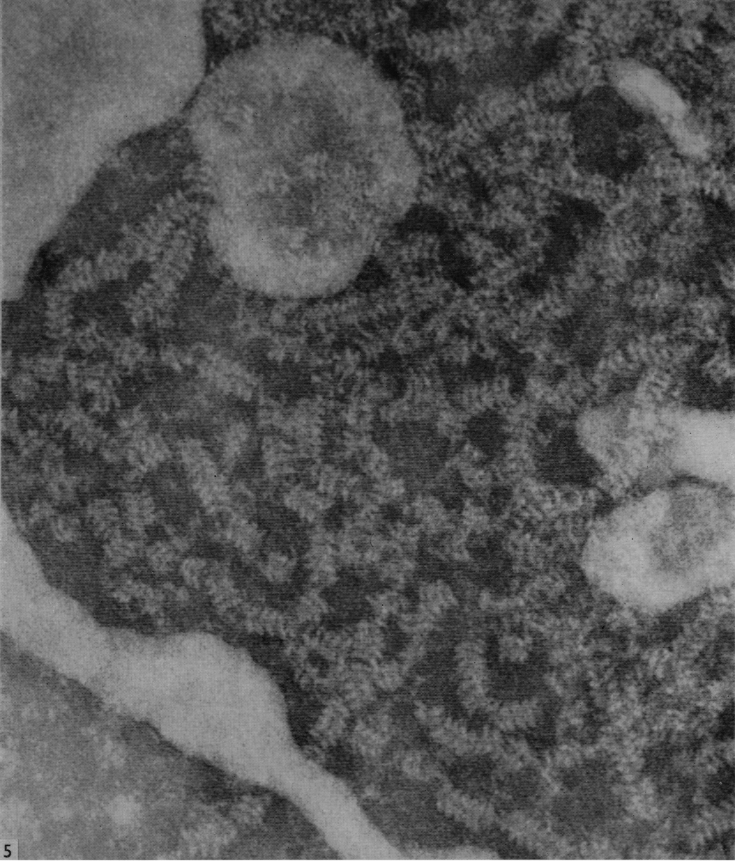
A single virus particle
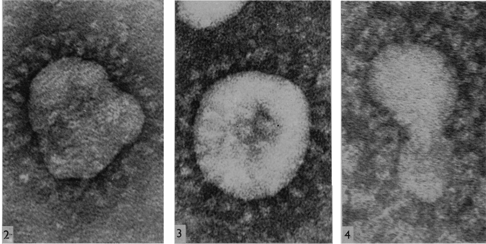
Their research article, titled ‘The morphology of three previously uncharacterised human respiratory viruses that grow in organ culture’, was published in JGV in 1967.
Images showing the distinct ‘fringe’ of protrusions around the virus particle
According to Dr Almeida, these then-uncharacterised virus particles were similar to those she had previously extracted from chickens with infectious bronchitis.
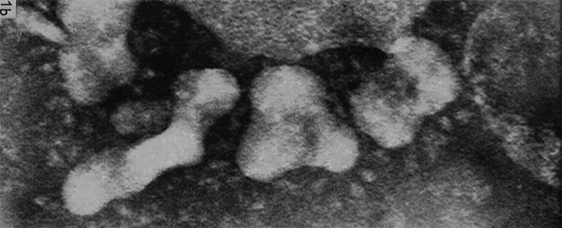
The authors described the ‘distinct layer of projections’ on the surface of the virus particles. Later, these projections, which appeared halo or crown-like under the microscope, inspired the name (corōna is the Latin for crown, or wreath).
The full article is fully Open Access and free to read on the JGV website.