Visualising bluetongue virus in midges
Posted on March 24, 2016 by Anand Jagatia
Today at the Society’s Annual Conference, Alice Muntzer spoke about a new way to visualise virus particles inside biting midges that spread disease.
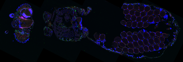
This unusual looking image is a fluorescently labelled section of a Culicoides biting midge. On the left you can see the insect’s head and compound eyes. On the right of the image is the midge’s abdomen, which is full of eggs.
Culicoides biting midges are known to spread diseases like bluetongue (which infects animals like sheep, cattle and goats) and African horse sickness (which affects horses but also mules and donkeys). Both diseases can cause high mortality and generate large economic costs, so they are of particular concern to owners.
For infected midges to transmit the virus, it has to spread through its tissues to the salivary glands, but the insect has a number of internal “barriers” that limit viral infection. In the midge, the barriers are found in the midgut and inhibit the virus from entering and leaving this area.
Understanding these barriers is important, because they determine whether the virus is able to infect the salivary glands, and therefore if the midge is capable of transmitting virus to another host. But scientists don’t currently know very much about how viruses are disseminated within midges or other disease vectors like mosquitoes.
Alice Muntzer is a researcher at The Pirbright Institute, working on localising bluetongue virus inside Culicoides midges. Her group has developed a method for visualising and quantifying the virus, which can be seen in the image below.
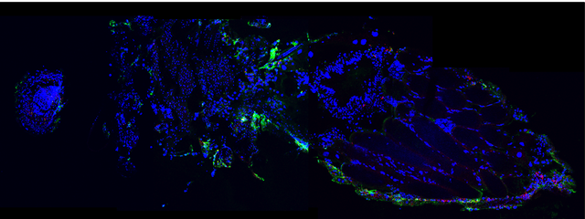
Using fluorescently-labelled antibodies that bind to the virus (shown in green in the above photo), the team has been able to show the tissues that contain bluetongue virus particles. More than that, they can also see the parts of the insect where the virus is actively replicating (shown in red). This was done by creating fluorescent tags that bind to genes that encode one of the viral proteins. These red tags only bind when the gene is expressed, or turned on, which happens during virus replication.
The method has allowed the team to track dissemination of the virus in whole midges for the first time. In the image above, you can see that virus is present in the compound eye, the brain, and the midgut.
“Initially I looked at a time series of infection in the midges,” says Alice. “Looking at which tissues become infected and how much virus you get in certain areas at different times, because we don’t even know the answers to basic questions like this.”
Most studies in the past have relied on dissecting out the midgut or the salivary glands and testing these for presence of the virus, but these methods are prone to cross-contamination. Alice’s technique also gives more information and context about infection within the whole insect.
The team was able to see whether some midges were completely uninfected and whether infection phases varied between different individuals. They have also used the system to look at the impact of external factors on the infection progression, like the amount of virus ingested by a midge in a blood meal, or the external temperature, which regulates insect body temperature.
“This has been a great model for looking in detail at the process of infection, and testing it under different parameters,” says Alice. “It lets us see when the salivary gland becomes infected, and by looking at different conditions we can see when more midges will develop infection and therefore when there will be more transmission.”