Jam Talks: Microbes and Medicine
Posted on June 26, 2019 by Liam Rooney
The May 2019 Junior Awards for Microbiology (JAM) talks took place in Birmingham on the 10 May. PhD student Liam Rooney was speaking, and in the third of our blogs from speakers at the JAM talks, Liam takes us through his research.
Having the privilege of working at the interface of biology and physics is something that makes every day exciting. By finding new applications for advanced microscopy in the field of microbiology I get to observe things which no one else has seen before; from how bacteria come together to organise in large communities, to how microbes move and swarm in flocks to seek out nutrients and prey. My research spans several areas within microbiology, all with the common theme of adapting novel or under-appreciated microscopy techniques to the field.
The optical, or light microscope was first developed back in the 17th century and pioneered by figures such as Galileo Galilei, Antoni Van Leeuwenhoek and Robert Hooke. Since then the microscope had been steadily developed, but the technology has jumped forward in the last 30 years with the introduction of super-resolution techniques and computational analysis tools. These new tools are fantastic for studying eukaryotic cells, however individual bacteria are so small they are incredibly hard to image, making it difficult to study bacterial dynamics. We therefore need to start using new microscopy techniques if we want to look at novel behaviours in live bacteria.
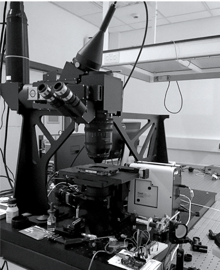
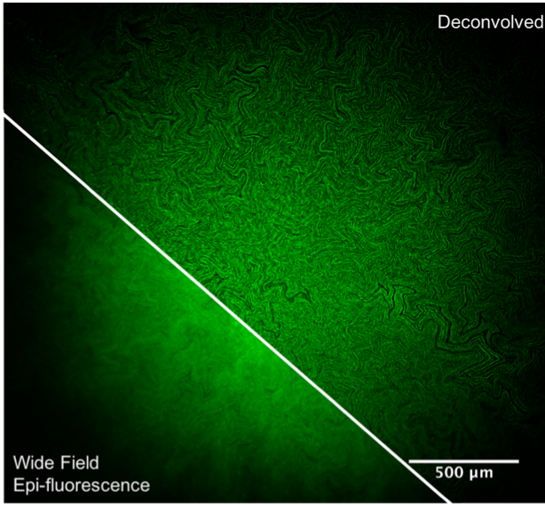
The first aspect of my work involves studying bacterial biofilm organisation. Biofilms are communities of micro-organisms that stick to each other and to surfaces using self-secreted extracellular polymeric substance (EPS), a sticky slime-like material consisting of DNA, polysaccharides and proteins. We study the internal architecture of bacterial biofilms using a microscope called the Mesolens. This microscope has the huge advantage of being capable of capturing images of large samples with high resolution without the need for additional processing. The Mesolens is the most radical redesign of the optical microscope in over 100 years, and is able to capture high spatial resolution over large scales due to the unique combination of a lens with low magnification and high numerical aperture (Figure 1). Using the Mesolens we have discovered channels within Escherichia coli biofilms. These channels have no bacteria or EPS within them and we believe they are used for nutrient uptake (Figure 2). Biofilms are a huge health issue as they protect the bacteria from outside factors such as antibiotics. However, these channels could be used to ‘feed’ the bacteria with biofilm disrupters, making the bacterial infection easier to treat.
Another aspect of my research is bacterial motility, where we study the gliding behaviour of myxobacteria. The motility mechanisms of Myxococcus xanthus have been well-documented using conventional microscopy methods. However, thus far only the gliding behaviours from a birds-eye view (i.e. the lateral dynamics) have been observed. We have used a technique called Interference Reflection Microscopy (IRM) which allows us to look at how the cells behave in 3D as they glide. This technique uses reflected light to generate, contrast and build an image. The reflected light creates a fringe pattern which acts as a contour map of the cell, just like the orange contour lines on an Ordinance Survey map. Using IRM, we can observe changes in the height of the cell as it glides down to 90 nm and have revealed that M. xanthus undulates as it moves and that the height of the cell along the body changes as they glide.
More recently I’ve been creating a soil-like culture environment for soil bacteria that we can use under an optical microscope. To achieve this, we have built a transparent soil-based platform in which bacteria can be grown and imaged without affecting their normal behaviour. The aim here is to determine whether growing bacteria in a 3D environment, similar to what would be found in nature, can alter the interactions between different groups of bacteria and their production of specialised metabolites (e.g. antibiotics).
Having such a varied and interdisciplinary PhD project has exposed me to a number of opportunities and has allowed me to develop a whole host of technical skills along the way. Getting the chance to attend both microscopy and microbiology-focused meetings and to present my research has been a great confidence builder and broadened my network of colleagues and collaborators. This included speaking at this year’s JAM Talks in Birmingham, where I was able to showcase my work in a friendly and supportive environment. The relaxed atmosphere of this student-led initiative meant I came away with some great experimental suggestions and new insights on my research. I cannot recommend enough for any PhD students to apply for the next intake of JAM Talks and to take the chance to share their work with other early career researchers.
The Junior Awards for Microbiology (JAM) talks is a monthly junior seminar series aimed at integrating and connecting young researchers around Europe.
To learn more about the JAM talks, listen to our podcast with Alice Lanne and Anja Djokic, two members of the organising team, or read the blog written by Daniel Morse, who spoke at the talks in October.