Observing fungi in a Petri dish
Students should examine cultures in containers, which have been taped and closed. Colony morphology is a method that scientists use to describe the characteristics of an individual colony of fungi growing on agar in a Petri dish. It can be used to help to identify them.
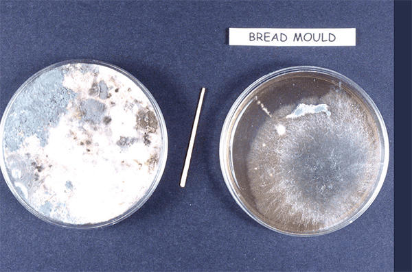
A circular piece of bread that has been allowed to go mouldy.
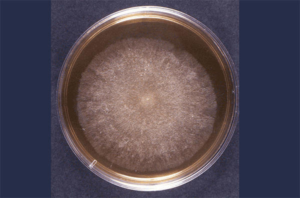
Plate 1 contains a circular piece of bread that has been allowed to go mouldy. There is overgrowth on the plate and many different mould species can be seen. In between the two plates is a toothpick which was used to isolate the spores from one of the moulds from plate 1. The mould spores from the toothpick were inoculated onto malt extract agar, plate 2. Use the diagrams on colony morphology to help you interpret plate 2.

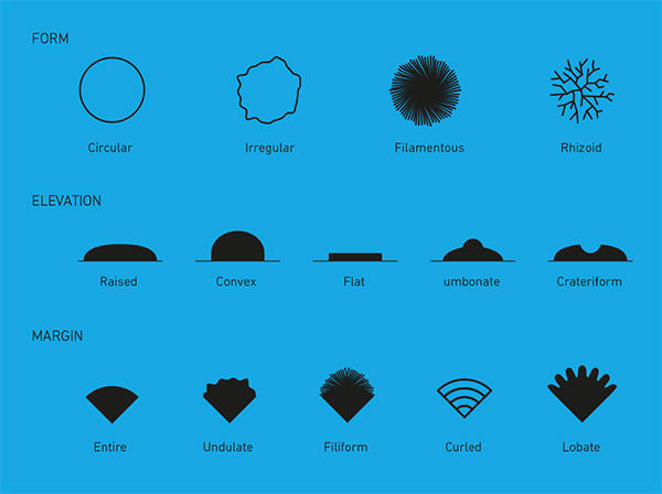
Different types of fungi will produce different-looking colonies, some colonies may be coloured, some colonies are circular in shape, and others are irregular. A specific terminology is used to describe common colony types. These are:
- Form – what is the basic shape of the colony? For example, circular, filamentous, etc.
- Size – the diameter of the colony. Tiny colonies are referred to as punctiform
- Elevation – this describes the side view of a colony. Turn the Petri dish on end.
- Margin/border – the edge of a colony. What is the magnified shape of the edge of the colony?
- Surface – how does the surface of the colony appear? For example, smooth, glistening, rough, wrinkled, or dull.
- Opacity – for example, transparent (clear), opaque, translucent (like looking through frosted glass), etc.
- Colour (pigmentation) – for example, white, buff, red, purple, etc.
Yeast colonies are very similar to bacterial colonies.
Moulds often have fuzzy edges. They usually turn into a different colour, from the centre outwards.